







The Gut–Vagus–Brain Loop: Microbiome, Inflammation, and Mood Regulation
The Gut–Vagus–Brain Loop: Microbiome, Inflammation, and Mood Regulation
The gut–vagus–brain loop describes the bidirectional communication between the gut microbiome, the vagus nerve, and the central nervous system (CNS). This complex network integrates neural, hormonal, and immune signalling and links digestion and microbial activity with emotional and cognitive processes. It underpins much of the current understanding of the “gut–brain axis”, illustrating how the state of the gut can directly influence mood, behaviour, and even mental health disorders.
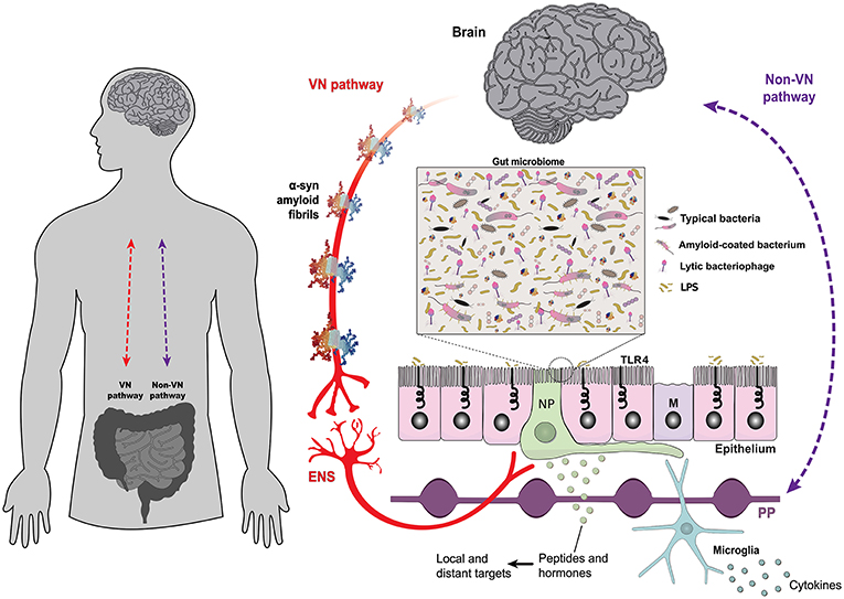
The bidirectional interplay between the brain and the gut is mediated by neural, such as the vagus nerve (VN-gateway), and humoral pathways, such as the lymphatic tissue and the bloodstream (Non-VN gateways). A monolayer of epithelial cells separates the intestinal lumen and the complex gut microbiome from the underlying lymphoid and enteric nervous tissues. The structure of alpha-synuclein amyloid fibrils (PDB 2N0A) is based on atomic-resolution molecular data from NGL Viewer (19). Members of the gut microbiome and their extracellular compounds may trigger responses in the VN through enteroendocrine cells, which are contacted by vagus nerve terminals through specialised structures called neuropods (NP) (20). Microbial antigens can cross the gut epithelium through microfold cells, playing a central role in localised inflammatory responses [adapted from Bohórquez et al. (21)]. Toll-like receptors are microbe-sensing proteins, present in intestinal epithelial cells, mediating recognition of commensal bacteria from the harmful/inflammatory ones. ENS, enteric nervous system; M, microfold cells; NP, neuropods; PP, Peyer's patches; TLR4, Toll-like receptor 4; VN, vagus nerve.
Source: Santos, S. F., de Oliveira, H. L., Yamada, E. S., Neves, B. C., & Pereira, A. (2019). Neural and humoral pathways of gut–brain communication. Frontiers in Neurology, 10, 574.
The Microbiome’s Role in Signalling
The gut microbiome produces a vast array of metabolites, neurotransmitters, and signalling molecules that influence the vagal tone and brain activity.
- Neurotransmitters: Certain gut bacteria (e.g., Lactobacillus, Bifidobacterium) produce γ-aminobutyric acid (GABA), serotonin, and dopamine precursors, all of which affect emotional regulation.
- Short-chain fatty acids (SCFAs): Butyrate, acetate, and propionate—produced through fermentation of dietary fibre—modulate inflammation and strengthen the intestinal barrier, indirectly affecting vagal afferent signalling.
- Tryptophan metabolism: Microbial modulation of tryptophan availability affects serotonin synthesis in the enterochromaffin cells of the gut, impacting mood and sleep.
Enterochromaffin Cells
Enterochromaffin cells (EC) are specialised neuroendocrine cells found throughout the lining of the gastrointestinal tract, particularly in the small intestine. They play a vital role in gut–brain communication by producing and releasing serotonin (5-hydroxytryptamine, 5-HT): a neurotransmitter that regulates gut motility, secretion, and mood.
About 90% of the body’s serotonin is synthesised in these cells, not in the brain. When food or microbial metabolites stimulate EC cells, they release serotonin into the gut wall, where it activates vagal afferent fibres and local enteric neurones. This signalling influences both digestion and emotional regulation via the vagus nervous system and the central nervous system.
Disruption of EC cell function or serotonin equilibrium has been associated with disorders such as irritable bowel syndrome (IBS), depression, and anxiety, underscoring their critical role in the gut–vagus–brain axis.
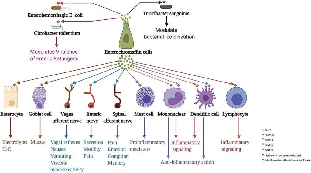
Enterochromaffin cells act on various neighbouring cells, neurons, and intestinal microbiota through the release of 5-HT. 5-HT acting on 5-HT4R stimulates epithelial cells to secrete electrolytes and H2O into the intestinal lumen and stimulates goblet cells to secrete mucus. Moreover, 5-HT acting on 5-HT3R of terminals of the spinal and vagus afferent nerves, and enteric nerves are involved in processes associated with emotion, cognition, memory, pain perception, distention, and chemical-evoked vagal reflexes, nausea, vomiting, visceral hypersensitivity, and intestinal functions, including secretion and motility. Furthermore, 5-HT acting on 5-HT1AR induces mast cell adhesion, migration, and degranulation. In addition, 5-HT acting on 5-HT7R in immune cells, including monocytes, lymphocytes, and dendritic cells, may be implicated in inflammation signalling. In contrast, 5-HT acting on 5-HT1AR in monocytes and dendritic cells has an anti-inflammatory role. In addition to these functions, 5-HT action on 5-HT receptors of enteric bacteria, sodium symporter-related protein of Turicibacter sanguinis, and membrane-bound histidine sensor kinase of enterohemorrhagic Escherichia coli and Citrobacter rodentium, plays a crucial role in modulating bacterial colonisation and virulence of enteric pathogens. 5-HT, serotonin; 5-HTR, serotonin receptor.
Source: Tao, E., Zhu, Z., Hu, C., Long, G., Chen, B., Guo, R., Fang, M., & Jiang, M. (2022). Potential roles of enterochromaffin cells in early life stress-induced irritable bowel syndrome. Frontiers in Cellular Neuroscience, 16, 837166.
The Vagus Nerve as a Communication Pathway
The vagus nerve is the principal neural route connecting the gut and the brain. Around 80%–90% of its fibres are afferent, transmitting sensory information from the viscera to the brainstem (particularly the nucleus tractus solitarius). From the NTS, interoceptive signals are relayed to the hypothalamus, amygdala, insula, and prefrontal cortex, integrating visceral state with emotional appraisal and cognitive regulation.
- Sensory input: The vagus nerve detects mechanical distension, gut hormones, and microbial metabolites.
- Efferent output: In response, it modulates motility, secretion, and immune responses through parasympathetic control.
- Anti-inflammatory reflex: Through the “cholinergic anti-inflammatory pathway”, efferent vagal fibres release acetylcholine, inhibiting the release of pro-inflammatory cytokines such as TNF-α and IL-6 from macrophages.
Macrophages
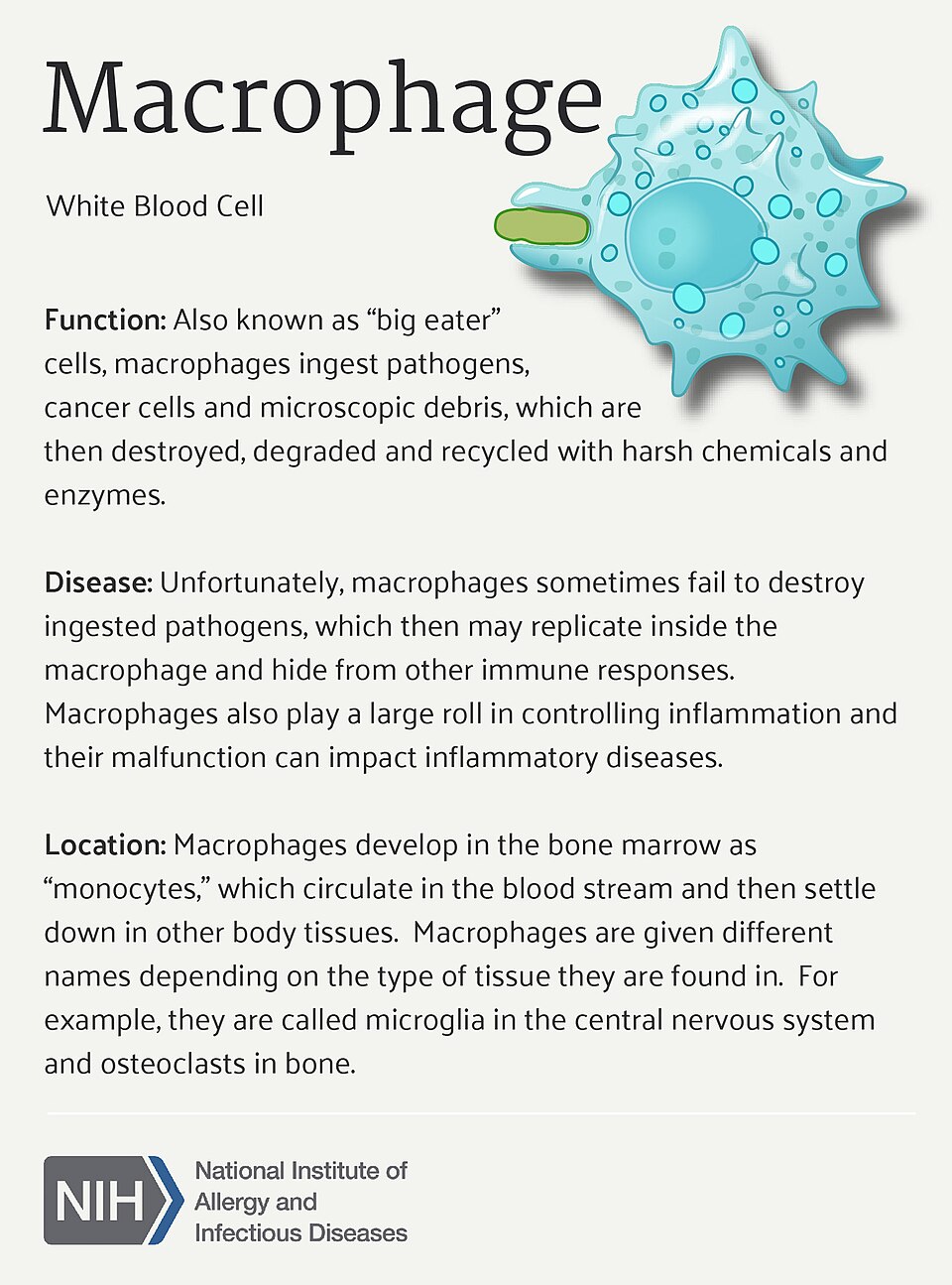
Macrophages are a type of white blood cell that form a crucial part of the body’s innate immune system. Found throughout tissues, including the gut, liver, lungs, and brain, they act as the body's “clean-up crew”, detecting, engulfing, and destroying pathogens, dead cells, and cellular debris.
In the gut, macrophages play an essential role in maintaining immune balance. They help protect the intestinal lining from infection while preventing excessive inflammation that could damage tissues.
Through the vagus nerve's cholinergic anti-inflammatory pathway, macrophage activity can be regulated by the acetylcholine released from efferent vagal fibres. This signalling helps to reduce the production of pro-inflammatory cytokines such as TNF-α, IL-1β, and IL-6 from macrophages.
When macrophages become overactivated (for example, in response to dysbiosis or a “leaky gut”) they contribute to chronic inflammation, which is implicated in many conditions, including IBS, depression, and autoimmune disorders. Balanced macrophage activity is therefore vital for both gut health and emotional regulation via the gut–vagus–brain axis.
Inflammation and Immune Cross-Talk
Chronic low-grade inflammation is increasingly recognised as a mediator between gut dysbiosis and mood disorders, such as depression and anxiety.
- Dysbiotic microbiota can increase intestinal permeability (“leaky gut”), allowing lipopolysaccharides (LPS) to enter circulation.
- These LPS molecules trigger systemic immune activation, raising cytokine levels that influence the CNS via both humoral routes and vagal afferent signals.
- Elevated cytokines can alter neurotransmitter metabolism, reduce neurogenesis, and impair the function of the hypothalamic–pituitary–adrenal (HPA) axis.
Lipopolysaccharides (LPS)
Lipopolysaccharides (LPS) are large molecules found in the outer membrane of Gram-negative bacteria. They are sometimes referred to as endotoxins because of their powerful ability to trigger immune responses in the human body.
Under normal conditions, LPS remains contained within the gut lumen. However, when the intestinal barrier becomes compromised, a condition often called “leaky gut”, LPS can pass into the bloodstream. Their presence activates immune cells such as macrophages, which release pro-inflammatory cytokines, including TNF-α and IL-6.
This immune activation contributes to systemic inflammation, which can affect brain function and mood through the gut–vagus–brain axis. Elevated LPS levels have been associated with fatigue, low mood, and metabolic disorders. Maintaining a healthy gut microbiome and a strong intestinal barrier helps prevent LPS translocation and reduce inflammation-related stress on both the body and mind.
Gram-Negative and Gram-Positive Bacteria
Bacteria are broadly classified as Gram-negative or Gram-positive based on differences in their cell wall structure, identified by the Gram staining method developed by Danish bacteriologist Hans Christian Gram in 1884. This distinction is essential in microbiology, medicine, and gut health research, as it influences how bacteria interact with the immune system and respond to antibiotics.
Gram-Positive Bacteria: These bacteria have a thick peptidoglycan layer in their cell wall, which retains the violet dye during Gram staining, making them appear purple under a microscope. They lack an outer membrane. Examples include Lactobacillus, Streptococcus, and Clostridium. Many Gram-positive species in the gut are beneficial, aiding digestion and maintaining intestinal barrier integrity.
Gram-Negative Bacteria: These bacteria have a thin peptidoglycan layer but possess an additional outer membrane that contains lipopolysaccharides (LPS). During Gram staining, they do not retain the violet dye and instead appear pink or red. Examples include Escherichia coli (E. coli), Salmonella, and Helicobacter pylori. The LPS in their outer membrane can trigger strong immune and inflammatory responses if it enters the bloodstream.
In a balanced microbiome, both Gram-positive and Gram-negative bacteria coexist in harmony. However, when gut dysbiosis occurs, an overgrowth of Gram-negative bacteria may increase LPS exposure, contributing to inflammation and disturbances in the gut–vagus–brain axis.
Mood Regulation and Emotional Processing
Emerging research links alterations in gut microbiota composition and vagal tone with changes in emotional regulation:
- High vagal tone is correlated with better emotional resilience, reduced anxiety, and improved social engagement (as described in Polyvagal Theory).
- Low vagal tone and dysbiosis are associated with stress vulnerability, depressive symptoms, and cognitive slowing.
- Interventions such as probiotics, vagus nerve stimulation, mindfulness, and IEMT may help restore balance in the gut–vagus–brain network by enhancing parasympathetic regulation.
Understanding this dynamic loop emphasises the importance of treating the gut as an integral component of emotional and psychological well-being, with direct influence on autonomic tone, limbic reactivity, and prefrontal regulation. Healthy microbial diversity, an effective vagal tone, and balanced immune signalling form the physiological foundation for mood stability and the adaptive stress response.