







The Autonomic Nervous System
Overview
The autonomic nervous system (ANS) governs the involuntary functions of the body that maintain physiological balance and survival. It regulates processes such as heart rate, blood pressure, respiration, digestion, and glandular secretion and indirectly influences immune function through neuroendocrine and inflammatory pathways. Unlike the somatic nervous system, which controls voluntary movement, the autonomic system operates largely outside conscious awareness, adjusting the body’s internal state moment to moment.
The ANS is a core interface between emotional experience and physiological expression. Changes in mood, threat perception, or attention are mirrored by shifts in heart rhythm, breathing, muscle tone, and visceral activity. For practitioners of IEMT, understanding these mechanisms provides a framework for recognising how the rapid emotional changes observed in sessions correspond to underlying autonomic adjustments.
The Somatic Nervous System
Overview:
The somatic nervous system controls voluntary movements and transmits sensory information from the body to the central nervous system. It enables conscious interaction with the environment through skeletal muscles, allowing coordinated actions such as walking, speaking, and manipulating objects.
Physiological Role:
This system consists of motor neurones, which transmit signals from the brain and spinal cord to muscles, and sensory neurones, which convey information from the skin, joints, and muscles back to the brain. Unlike the autonomic system, somatic activity is under conscious control. However, both systems operate in close coordination, voluntary movement often triggers autonomic adjustments such as changes in heart rate, respiration, or blood flow to muscles.
Insular Cortex
Main Divisions of the ANS
The autonomic nervous system is traditionally divided into three components, the sympathetic, the parasympathetic, and the enteric systems. These operate together to maintain internal stability while responding to environmental demands.

- Sympathetic nervous system (SNS): Activates the body’s resources in response to stress or challenge. It increases heart rate, dilates the pupils, redirects blood to muscles, and releases adrenaline and noradrenaline. This system underpins the classic “fight or flight” response.
- Parasympathetic nervous system (PNS): Promotes relaxation, digestion, and recovery. It slows the heart rate, stimulates salivation and digestive activity, and supports restorative processes. The vagus nerve is the principal parasympathetic conduit connecting the brain and body.
- Enteric nervous system (ENS): Sometimes called the “second brain”, this network of neurones embedded in the gastrointestinal tract regulates digestion independently but remains closely linked to both sympathetic and parasympathetic activity. It communicates extensively via the vagus nerve, contributing to the gut–brain axis.
Autonomic Balance and Flexibility
Healthy autonomic function depends on the body’s ability to shift fluidly between states of activation and recovery. The sympathetic and parasympathetic branches are not opposites but complementary systems that maintain adaptive responsiveness. In everyday life, this balance allows for alertness, social engagement, and rest without prolonged stress or exhaustion.
When one branch becomes chronically dominant (such as sympathetic overdrive or parasympathetic withdrawal), the result can be emotional instability, fatigue, or physiological dysregulation. Patterns of chronic anxiety, hypervigilance, or dissociation often reflect these enduring autonomic imbalances. Recognising them enables practitioners to frame such conditions as adaptive physiological patterns rather than fixed psychological traits.
The Psychology and Causation of Hypervigilance
Hypervigilance refers to a sustained state of heightened sensory sensitivity and threat monitoring. It is a psychological and physiological situation in which the individual’s attention system becomes chronically biased toward detecting danger, even in safe environments.
From a neurobiological perspective, hypervigilance reflects persistent activation of the amygdala and limbic threat circuits, often coupled with reduced prefrontal regulation. The hypothalamic–pituitary–adrenal (HPA) axis remains overactive, leading to elevated levels of stress hormones such as cortisol and adrenaline. This leads to constant scanning, startle reactions, sleep disturbance, and difficulty concentrating.
Psychologically, hypervigilance commonly develops after trauma, chronic stress, or attachment disruption, where vigilance once served an adaptive protective role. Over time, however, the pattern becomes self-reinforcing, the person interprets neutral cues as potential threats, which confirms their sense of danger, perpetuating the loop.
Therapeutically, interventions focus on downregulating autonomic arousal and retraining attention and interoceptive awareness, often through body-based methods, trauma-focused therapy, mindfulness, or parasympathetic-oriented regulation practices such as breath-based and body-focused methods.
Central Control of the ANS
The autonomic nervous system is coordinated by several brain regions, including the hypothalamus, amygdala, insular cortex, and brainstem nuclei such as those within the medulla oblongata. These centres integrate sensory information, emotional appraisals, and environmental cues to maintain homeostasis.
The hypothalamus acts as the principal regulator, balancing endocrine, immune, and autonomic outputs. The amygdala interprets emotional significance and can trigger sympathetic activation under threat. The insula contributes interoceptive awareness, the perception of internal bodily states, which influences conscious emotional experiences. Collectively, these systems determine the physiological signature of emotion.
The Insular Cortex
Overview:
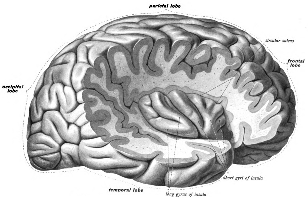
The insular cortex, or insula, is a region of the brain located deep within the lateral sulcus, where the frontal, temporal, and parietal lobes meet. It serves as a hub for integrating sensory, emotional, and bodily information, linking subjective feeling states to physiological processes.
Physiological Role:
The insula receives interoceptive input from the viscera via the vagus nerve and other autonomic pathways. It processes sensations such as heartbeat, respiration, temperature, and visceral tension, contributing to awareness of internal bodily states. This information is then integrated with emotional and cognitive processing, shaping the subjective experience of feeling. The insula thus plays a key role in self-awareness, empathy, and the embodiment of emotion.
Homeostasis and Allostasis
Homeostasis refers to the body’s capacity to maintain stable internal conditions despite external change. The concept of allostasis expands on this by emphasising adaptive adjustments: the way the body actively anticipates and prepares for stressors. The autonomic system orchestrates these adjustments by modulating heart rate, blood pressure, hormonal output, and immune activity.
However, when demands are chronic or overwhelming, the cost of maintaining allostasis becomes high, leading to what is termed an “allostatic load”. This cumulative strain can impair vagal regulation, lower heart rate variability, and contribute to both physical and emotional exhaustion. Understanding this process helps explain how repeated stress exposure can lead to enduring emotional and physiological patterns.
Allostatic Load
Overview:
Allostatic load refers to the cumulative physiological strain placed on the body as it repeatedly adapts to stress. While short-term activation of the autonomic, endocrine, and immune systems supports survival, chronic or repeated stress responses can exhaust these mechanisms and disrupt balance.
Physiological Role:
During stress, the sympathetic nervous system and hypothalamic–pituitary–adrenal (HPA) axis release hormones such as cortisol and adrenaline to mobilise energy and maintain alertness. When these responses remain elevated or fail to deactivate properly, the body experiences wear and tear across multiple systems, including cardiovascular, immune, and metabolic functions. Over time, this persistent activation is associated with reduced vagal tone, lower heart rate variability, and increased vulnerability to illness and emotional dysregulation.
Interoception and Emotional Experience
Interoception (the perception of signals from within the body) plays a central role in emotion and self-awareness. The autonomic system generates much of this sensory input, transmitting information about heart rate, respiration, and visceral sensation to the brain via afferent fibres, primarily through the vagus nerve alongside important spinal sensory pathways.
Feelings such as calm, fear, or tension emerge partly from the brain’s interpretation of these bodily states. Autonomic regulation, therefore, forms the foundation of emotional life. By helping clients access awareness of internal states, therapeutic interventions can foster greater stability and self-regulation.
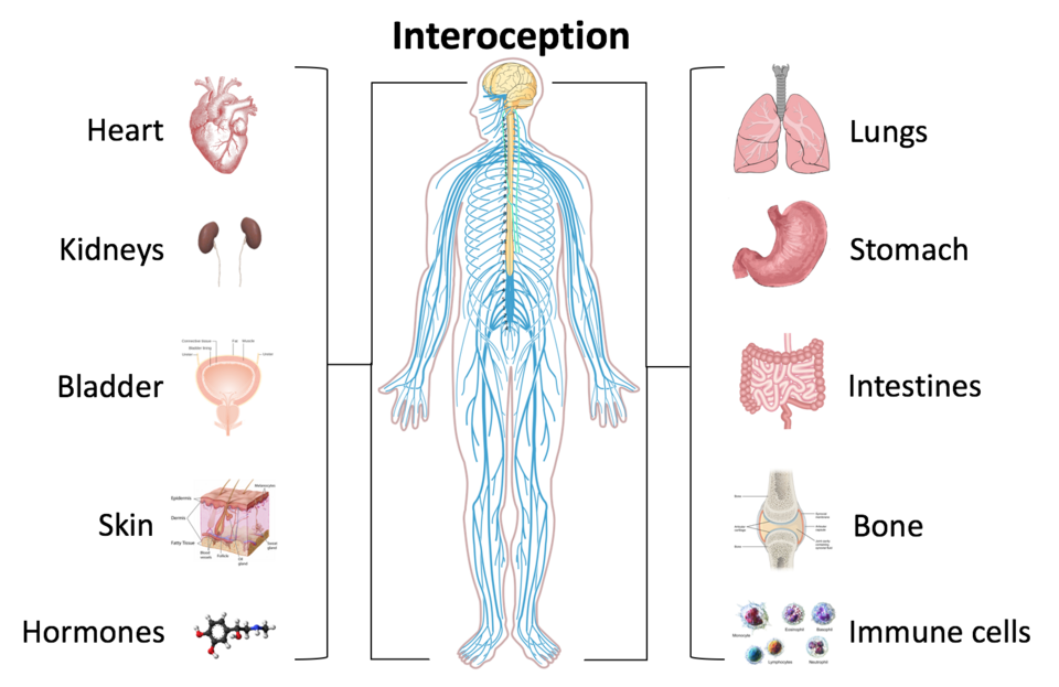
Interoception is a process involving the nervous system and innervated organs and organ systems. Organs involved in interoception are indicated on the right and left of the body.
Source: Schappelle. (2017, August 16). Interoception and the body [Illustration]

License: CC BY-SA 4.0.
Interoception
Interoception is the process by which the brain senses, interprets, and integrates signals originating from within the body. These signals include information about heart rate, respiration, temperature, hunger, thirst, and visceral sensations. Interoception allows the body to maintain internal stability while also forming the sensory foundation of emotional experience.
Clinical observation and research indicate that interoceptive awareness is often reduced in a significant proportion of individuals, leading to difficulty differentiating between epigastric discomfort and chest pain, or between emotional distress and cardiac symptoms.
Physiological Role:
Interoceptive signals are transmitted primarily through the vagus nerve and spinal pathways to brain regions such as the insular cortex, anterior cingulate cortex, and brainstem nuclei. This continuous feedback informs the brain about physiological needs and states, supporting adaptive regulation of emotion, attention, and behaviour. Accurate interoceptive processing contributes to self-awareness and emotional stability, while disturbances in interoception are linked to anxiety, alexithymia, and dissociative experiences.