







Depression and the Vagus Nerve
The relationship between the vagus nerve and depression has become a focal point in psychophysiological and neuropsychiatric research. The vagus nerve acts as a critical interface between the body’s internal organs and the brain, influencing immune signalling, autonomic regulation, inflammation, and mood. Emerging evidence suggests that dysfunction of vagal pathways may contribute to the neurobiological mechanisms underlying depressive disorders.
Neurobiological Foundations
Major depressive disorder (MDD) involves widespread changes in neural activity, neuroendocrine function, and inflammatory regulation. The vagus nerve plays a key modulatory role in these systems through both its afferent fibres (transmitting visceral sensory information to the brain) and efferent fibres (mediating parasympathetic control of target organs). Approximately 80% of vagal fibres are afferent, carrying signals from the heart, lungs, and gut to the nucleus tractus solitarius (NTS) in the brainstem.
Diagnostic Criteria for Major Depressive Disorder (MDD)
Major Depressive Disorder is diagnosed according to criteria outlined in the Diagnostic and Statistical Manual of Mental Disorders, Fifth Edition (DSM-5).
The condition requires the presence of five or more of the following symptoms during the same two-week period, representing a change from previous functioning, and at least one of the symptoms must be depressed mood or loss of interest or pleasure (anhedonia):
- Persistent depressed mood most of the day, nearly every day.
- Markedly diminished interest or pleasure in almost all activities.
- Significant weight loss or gain, or changes in appetite.
- Insomnia or hypersomnia.
- Psychomotor agitation or retardation observable by others.
- Fatigue or loss of energy.
- Feelings of worthlessness or excessive or inappropriate guilt.
- Diminished ability to think, concentrate, or make decisions.
- Recurrent thoughts of death, suicidal ideation, or suicide attempt.
Symptoms must cause clinically significant distress or impairment in social, occupational, or other important areas of functioning. They cannot be attributable to the physiological effects of a substance or another medical condition.
The diagnosis also specifies severity (mild, moderate, or severe) and episode pattern (single or recurrent) and may include specifiers such as melancholic features, atypical features, or anxious distress.
To understand how these psychological symptoms become biologically embedded in the body, it is essential to examine the role of the vagus nerve. The vagus nerve serves as the primary communication highway between the internal organs and the brain. Sensory information from the heart, lungs, and gut travels upward along vagal afferent fibres and converges in a key brainstem structure known as the Nucleus Tractus Solitarius (NTS). The NTS functions as a central relay station, translating bodily signals into neural messages that influence emotional regulation, stress responses, and mood.
The NTS projects to several mood-relevant structures, including the locus coeruleus (noradrenergic nucleus), dorsal raphe nucleus (serotonergic system), amygdala, hypothalamus, and prefrontal cortex. Through these circuits, the vagus nerve modulates neurotransmitters, stress hormones, and neuroinflammatory pathways that shape emotional and cognitive states.
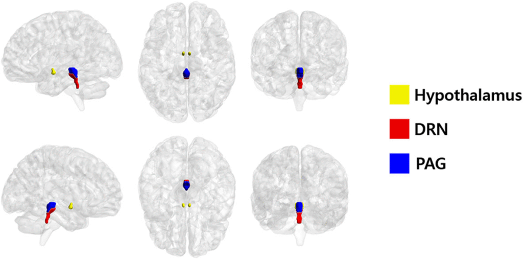
Dorsal Raphe Nucleus (DRN)
The dorsal raphe nucleus is a cluster of neurones located in the midline of the brainstem, within the pons and midbrain. It is the largest serotonergic nucleus in the brain and serves as the primary source of serotonin (5-HT) projections to widespread regions, including the prefrontal cortex, hippocampus, amygdala, and hypothalamus.
Functionally, the DRN plays a central role in mood regulation, sleep–wake cycles, stress response, and emotional behaviour. Altered activity or connectivity of the dorsal raphe has been linked to depression, anxiety, and stress-related disorders.
The DRN receives regulatory input from structures such as the nucleus tractus solitarius (via the vagus nerve), locus coeruleus, and prefrontal cortex, allowing it to integrate autonomic, sensory, and emotional information into serotonergic modulation of the brain.
The Inflammatory Pathway and Cytokine Signalling
Chronic low-grade inflammation has been strongly implicated in the pathophysiology of depression. Elevated pro-inflammatory cytokines such as interleukin-6 (IL-6), tumour necrosis factor-alpha (TNF-α), and interleukin-1β (IL-1β) have been found in subsets of depressed individuals. These cytokines can alter neurotransmitter metabolism, reduce synaptic plasticity, and disrupt neurogenesis, particularly in the hippocampus.
Interleukin-6 (IL-6)
Interleukin-6 is a pro-inflammatory cytokine produced by immune cells such as macrophages, T-cells, and microglia, as well as by endothelial and epithelial tissues in response to infection, injury, or psychological stress. It acts as a key mediator between the immune system and the central nervous system.
In the brain, IL-6 can cross the blood–brain barrier or be released locally by activated glial cells. Elevated IL-6 levels influence neurotransmitter metabolism, particularly serotonin and dopamine pathways, and contribute to neuroinflammation and reduced neuroplasticity, especially in the hippocampus.
High circulating IL-6 has been consistently associated with major depressive disorder, fatigue, and sickness behaviour, linking chronic inflammation to mood dysregulation. The vagus nerve, through the cholinergic anti-inflammatory pathway, helps suppress IL-6 release, highlighting a physiological bridge between emotional health and immune balance.
The vagus nerve exerts regulatory effects via the cholinergic anti-inflammatory pathway. When activated, efferent vagal fibres release acetylcholine, which binds to α7 nicotinic acetylcholine receptors on macrophages and microglia. This interaction inhibits the release of pro-inflammatory cytokines, helping to maintain immune homeostasis. Diminished vagal tone (a marker of reduced parasympathetic activity) is associated with impaired anti-inflammatory control and greater vulnerability to depressive symptoms.
Neurotransmitter and Brain Circuit Interactions
Vagal afferents influence monoaminergic nuclei, which are central to mood regulation:
- Serotonin (5-HT): The dorsal raphe nucleus receives direct input from the NTS. Enhanced vagal signalling increases serotonergic firing, which may improve affective stability and sleep regulation.
- Norepinephrine: The locus coeruleus, a key noradrenergic centre, integrates visceral feedback via the NTS. Vagal stimulation can normalise noradrenergic tone, counteracting the hyperarousal and stress sensitivity seen in depression.
- GABA and glutamate: Vagal pathways also influence the balance between inhibition and excitation in cortical and limbic regions, shaping neural oscillations associated with emotional regulation.
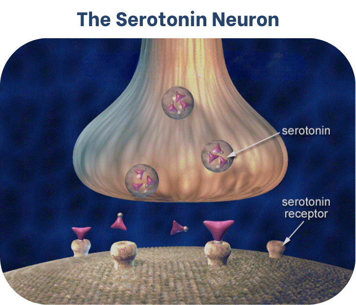
Source: National Institute on Drug Abuse. (2007). Neurobiology of ecstasy (MDMA): The serotonin neuron.
Neuroimaging studies of vagus nerve stimulation (VNS) have demonstrated increased activity and connectivity within the anterior cingulate cortex and orbitofrontal cortex, areas implicated in self-referential thought, emotional appraisal, and motivation.
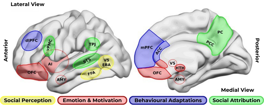
A simple classification of brain areas involved in social processing differentiates regions that participate in four related processes.
The first is the perception of basic social stimuli, such as biological motions (V5), parts of the body (extra-striate body area, EBA), and faces (fusiform face area, FFA).
Another process includes emotional and motivational appraisal, where the amygdala (AMY), the anterior insula (AI), the subgenual and perigenual anterior cingulate cortex (ACC), as well as the orbitofrontal cortex (OFC) participate.
These cortical structures are in interaction with subcortical structures as the ventral striatum (VS), and the hypothalamus (HTH).
These structures in turn interact with other regions which participate in the goal-directed, adaptive behaviors, and the categorization processes, such as the dorsolateral (dlPFC) and the medial prefrontal cortex (mPFC) and the ACC anterior cingulate cortex. Finally for social attribution, areas like the ventral premotor cortex (vPMC), the superior temporal sulcus (STS), the AI, the posterior cingulate cortex (PCC), and the precuneus (PC) participate in more automatic, bottom-up inferences of other people’s mental states; whereas structures like the mPFC and the temporo-parietal junction (TPJ) are involved in more cognitive theory of mind skills.
Billeke, P., & Aboitiz, F. (2013). Social cognition in schizophrenia: from social stimuli processing to social engagement. Frontiers in Psychiatry, 4(4).
Monoamines and Monoaminergic Nuclei
Monoamines are a class of neurotransmitters derived from aromatic amino acids that regulate mood, motivation, attention, and arousal. The main monoamines in the human brain are serotonin (5-HT), dopamine (DA), noradrenaline (norepinephrine, NE), and, to a lesser extent, histamine. They are called “monoamines” because each contains one amino group connected to an aromatic ring by a two-carbon chain.
Monoamines are synthesised in small, evolutionarily ancient clusters of neurones known as monoaminergic nuclei, located primarily in the brainstem and hypothalamus. Despite their limited number, these nuclei project widely throughout the brain, exerting diffuse and modulatory control over neural networks.
Key monoaminergic nuclei include:
- Dorsal and Median Raphe Nuclei: major sources of serotonin, influencing mood, sleep, and emotional regulation.
- Locus Coeruleus: primary centre for noradrenaline, involved in alertness, attention, and stress response.
- Substantia Nigra and Ventral Tegmental Area (VTA): dopaminergic nuclei governing reward, motivation, and motor control.
- Tuberomammillary Nucleus (Hypothalamus): primary histaminergic source, contributing to wakefulness and appetite regulation.
Dysfunction in monoaminergic systems, through altered synthesis, receptor sensitivity, or synaptic clearance, is strongly implicated in depression, anxiety, and other affective disorders. Many antidepressant medications act by enhancing monoaminergic transmission, such as selective serotonin reuptake inhibitors (SSRIs) or noradrenergic–dopaminergic reuptake inhibitors.
Autonomic Dysregulation and Stress
Depression often coincides with a state of autonomic imbalance, characterised by reduced parasympathetic (vagal) tone and heightened sympathetic activation. This manifests as low heart rate variability (HRV), elevated resting heart rate, gastrointestinal dysfunction, and sleep disturbances. Reduced vagal activity diminishes the body’s ability to recover from stress, maintaining chronic hypothalamic–pituitary–adrenal (HPA) axis activation and elevated cortisol levels. Persistent cortisol exposure contributes to hippocampal atrophy, impaired synaptic plasticity, and decreased neurogenesis: hallmark features of depressive pathology.
Vagus Nerve Stimulation (VNS) and Depression Treatment
Implanted vagus nerve stimulators were approved for treatment-resistant depression in the early 2000s following evidence of sustained remission in patients unresponsive to pharmacotherapy. These devices deliver intermittent electrical pulses to the left cervical vagus nerve, modulating afferent activity to central mood circuits.
Clinical and neuroimaging data suggest that chronic VNS enhances the functional connectivity of prefrontal–limbic pathways, increases cerebral blood flow in the orbitofrontal cortex, and upregulates noradrenergic and serotonergic tone. Functional MRI studies have also demonstrated VNS-induced normalisation of amygdala hyperactivity, a common finding in depression. While the antidepressant response develops gradually over several months, long-term follow-ups show durable benefits in a subset of patients.
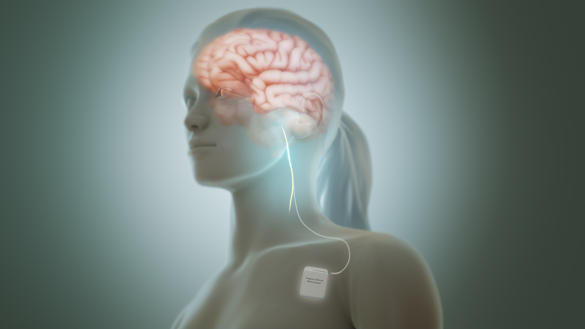
Source: Manu5. (n.d.). Vagus-nerve-stimulation. [Image] Scientific Animations.
License: CC BY-SA 4.0.
Non-Invasive Vagal Stimulation and Physiological Regulation
Recent research has explored transcutaneous vagus nerve stimulation (tVNS), which targets the auricular branch of the vagus nerve at the ear (typically the cymba conchae or tragus). Studies have reported reductions in depressive symptoms, improved HRV, and decreased inflammation markers. Although results are mixed and depend on stimulation parameters, tVNS represents a promising adjunctive or preventive approach for mood regulation.
Practical Applications: Everyday Strategies to Enhance Vagal Tone
While clinical interventions like transcutaneous vagus nerve stimulation (tVNS) show promise, accessible lifestyle practices can also support vagal function as complementary tools in depression management. Techniques such as diaphragmatic breathing (inhaling deeply for 4-6 seconds while expanding the abdomen, then exhaling slowly) activate the parasympathetic response, increasing heart rate variability (HRV) and dampening inflammatory cytokines like IL-6. Similarly, cold exposure (e.g., ending showers with 30 seconds of cool water) or humming/singing engages the vagus nerve via laryngeal branches, fostering a sense of calm and emotional resilience.
Hatha yoga and mindfulness meditation, which emphasise slow, rhythmic movements and breath awareness, have been linked to sustained improvements in vagal tone, with a 2022 meta-analysis in Psychosomatic Medicine reporting moderate effect sizes for reducing depressive symptoms in non-clinical populations. These practices are low-barrier entry points, ideal for integration into therapy or self-care routines, though they should complement, not replace, professional treatment. By cultivating daily habits that "tone" the vagus, individuals may experience subtle shifts in mood stability and stress recovery, aligning with the bidirectional gut-vagus-brain axis discussed earlier.
Clinical Caution: Breathing practices, cold exposure, and vagal stimulation techniques should be approached with care in individuals with cardiovascular disease, epilepsy, fainting disorders, pregnancy, or implanted medical devices. These practices are supportive wellness strategies and should not replace medical or psychological treatment.
Gut–Vagus–Brain Interactions
The gut microbiome influences vagal signalling through the production of short-chain fatty acids, tryptophan metabolites, and microbial neuroactive compounds. Dysbiosis can impair vagal afferent feedback and increase systemic inflammation, reinforcing depressive physiology. Conversely, enhancing vagal tone through dietary, breathing, and behavioural interventions may support gut–brain communication and physiological stress regulation, although direct causal effects on depressive disorders remain under investigation.
Summary of Key Mechanisms
| Mechanism | Effect on Depression | Vagal Involvement |
|---|---|---|
| Inflammatory cytokine signalling (IL-6, TNF-α, IL-1β) | Promotes neuroinflammation and disrupts neurotransmission | Inhibited via cholinergic anti-inflammatory pathway |
| Reduced HRV and parasympathetic tone | Associated with chronic stress and poor emotion regulation | Improved by vagal stimulation and breathing regulation |
| HPA axis hyperactivity | Elevated cortisol, hippocampal atrophy | Normalised through vagal afferent modulation of hypothalamus |
| Altered monoaminergic activity | Serotonin and noradrenaline imbalance | Vagal inputs influence raphe and locus coeruleus firing |
Emerging Research and Future Directions
Recent advancements in depression research are extending understanding beyond traditional monoaminergic models through the integration of neuroimaging, autonomic biomarkers, and psychotherapeutic outcome studies. For instance, the large-scale CoBalT randomised controlled trial, published in The Lancet Psychiatry (n = 469), investigated the addition of high-intensity cognitive behavioural therapy (CBT) to usual care (including antidepressant medication) for treatment-resistant depression. After six months, 46% of patients receiving adjunctive cognitive behavioural therapy (CBT) met the criteria for clinical response (defined as a ≥50% reduction in depressive symptoms), in contrast to 22% in the usual-care-only group. This research highlights the importance of multimodal intervention strategies for complex depressive presentations.
In parallel, mechanistic neuroimaging studies examining vagal afferent pathways demonstrate modulation of nucleus tractus solitarius projections to the amygdala, insula, and prefrontal cortex. These findings suggest a role for vagal signalling in emotional regulation, stress responsivity, and the neural circuits implicated in rumination and affective instability. Such data support growing interest in vagus-targeted interventions as adjunctive tools in mood disorder treatment.
Polyvagal theory, proposed by Stephen Porges, offers a theoretical framework suggesting that social engagement behaviours, such as prosodic vocalisations, facial expression, and safe eye contact, may influence autonomic state regulation via ventral vagal pathways. While this model is widely applied in trauma-informed and somatic therapeutic approaches, it remains debated within mainstream neuroscience due to limited direct anatomical and neurophysiological validation. It is therefore best regarded as a conceptual framework rather than an established mechanistic model of depression.
Future research directions include HRV-guided precision stimulation protocols, improved individualisation of non-invasive vagal stimulation parameters, and experimental investigations into gut–vagus–brain interactions. Although the gut microbiome is increasingly recognised as a contributor to affective regulation, microbiome interventions such as faecal microbiota transplantation currently remain restricted to research settings and are not approved clinical treatments for depressive disorders.
Collectively, these developments underscore the vagus nerve’s potential role as a key integrative regulator of emotional and physiological states. However, large-scale longitudinal trials remain necessary to optimise stimulation protocols, establish long-term safety, and personalise interventions across diverse populations, including adolescents and individuals with comorbid anxiety disorders.
Clinical and Research Implications
The vagus nerve’s role in depression exemplifies the growing understanding that mental health cannot be isolated from systemic physiology. Interventions that enhance vagal tone - whether electrical, behavioural, or somatic - may help restore autonomic balance, reduce neuroinflammation, and support emotional resilience. Ongoing research continues to refine the neural circuits and stimulation protocols that produce the most consistent therapeutic effects.
Integration with Integral Eye Movement Technique (IEMT): A Multimodal Approach
Integral Eye Movement Technique (IEMT) offers a novel lens for addressing vagus-mediated aspects of depression by targeting the sensory-motor patterns that reinforce negative self-schemas and autonomic imbalance. IEMT's eye-patterning exercises (guided shifts in gaze to reprocess emotional imprints) may indirectly influence autonomic regulation by modulating arousal processes associated with the locus coeruleus and by fostering interoceptive awareness, in a manner broadly analogous to how vagal afferents signal bodily states to the insula.
Preliminary observations from IEMT practitioners suggest that pairing these techniques with vagal-enhancing breathwork may accelerate shifts in affective states, potentially through co-regulation of oculomotor activity and parasympathetic engagement. For example, during an IEMT session, clients might alternate rapid eye saccades (to challenge cognitive distortions) with slow, vagotonic exhales, creating a therapeutic “window” that may support emotional flexibility and regulatory change. This effect may reflect modulation of emotion-regulation networks and serotonergic signalling rather than direct structural rewiring and therefore remains a hypothesis requiring formal experimental validation.
At present, these clinical observations should be interpreted as practice-based insights rather than established neurobiological mechanisms, highlighting the need for controlled psychophysiological and neuroimaging research to formally evaluate these effects.