






The Human Biome · Article 13 of 26
Histamine Intolerance & Mast Cell Activation – The Immune–Microbiome Connection
Educational and scope notice
This material is for education only and does not provide medical diagnosis or treatment advice. IEMT practitioners should remain within their professional scope and refer clients to an appropriately qualified healthcare professional where medical assessment, treatment, prescribed medication, supplements or restrictive diets are involved.
Histamine intolerance and Mast Cell Activation Syndrome (MCAS) are increasingly recognised as overlapping conditions involving immune dysregulation, inflammation, and altered microbiome balance. Though often discussed separately, both reflect the body’s difficulty in managing histamine: a naturally occurring compound involved in digestion, immunity, and the stress response.
What is Histamine?
Histamine is a signalling molecule (biogenic amine) that plays multiple roles in the body:
- Acts as a neurotransmitter in the brain, influencing wakefulness and mood.
- Regulates stomach acid secretion and intestinal motility.
- Functions as a key mediator of inflammation, helping immune cells respond to injury or infection.
Under normal conditions, histamine levels rise and fall as needed. However, when histamine accumulates faster than it can be broken down, symptoms develop, such as flushing, headaches, hives, nasal congestion, anxiety, or gastrointestinal discomfort. This state is referred to as histamine intolerance.
| Histamine Receptor | Primary Locations | Physiological Effects | Clinical Relevance | Gut–Brain Emotional Links |
|---|---|---|---|---|
| H₁ Receptor | Smooth muscle (airways, intestines) Endothelium CNS | Bronchoconstriction Vasodilation ↑ vascular permeability Wakefulness Allergic responses | Mediates allergic symptoms (itching, hives, rhinitis) Target for non-sedating antihistamines (e.g., cetirizine, loratadine) | Central H₁ tone promotes arousal Peripheral H₁ activation in the gut may drive visceral hypersensitivity and nausea. High histamine can exacerbate anxiety, irritability, and sleep disruption H₁ blockade is often sedating and anxiolytic. |
| H₂ Receptor | Gastric parietal cells Heart Uterus Vascular smooth muscle | Stimulates gastric acid secretion ↑ heart rate/contractility Vasodilation | Target for acid-reducing drugs (famotidine) in reflux/ulcer disease | Gastric acid affects microbial composition & nutrient availability, indirectly influencing mood via the gut–brain axis Some report reduced “histamine reactivity” with H₂ blockers May ease reflux-related sleep disturbance and autonomic arousal |
| H₃ Receptor | CNS & peripheral nerves (presynaptic) | Autoreceptor/heteroreceptor modulating release of histamine, dopamine, acetylcholine, serotonin; Influences arousal, attention, appetite | Experimental target for sleep disorders, ADHD, cognitive dysfunction | Key regulator of arousal and cognitive flexibility Altered H₃ signalling may contribute to fatigue, “brain fog”, appetite shifts, and stress responsivity Neural modulation can shape vagal tone and gut motility via autonomic pathways. |
| H₄ Receptor | Bone marrow Immune cells (mast cells, eosinophils, T-cells) GI tract | Immune cell chemotaxis Inflammatory signalling Mast-cell activity | Emerging target in IBD, asthma, autoimmune/inflammatory conditions | Bridges immune–neural crosstalk: mast-cell activation in the gut can affect permeability, microbiota, and cytokine signals reaching the brain, influencing anxiety-like behaviour and stress sensitivity; Potential role in IBS and neuroinflammation. |
| Common triggers: fermented foods, alcohol, aged cheese, processed meats, stress, oestrogen fluctuations, certain gut bacteria. Typical symptoms: flushing, headache, nasal congestion, digestive upset, anxiety, insomnia, and skin irritation. Note: While H₁ and H₂ receptors are well characterised, evidence for H₃ and H₄ involvement in emotional regulation and gut–brain signalling derives mainly from emerging translational and human observational studies. Further clinical research is ongoing. | ||||
Histamine and the Gut–Brain Connection
Histamine acts not only as an immune mediator but also as a neurotransmitter that influences mood, alertness, and stress reactivity. Within the gut, histamine released by mast cells and enterochromaffin-like cells communicates with the brain through several routes:
- Vagal signalling: Histamine activates receptors on the vagus nerve, transmitting information from the gut to brainstem centres involved in emotion and autonomic balance.
- Blood–brain communication: Although histamine itself crosses the blood–brain barrier poorly, it stimulates cytokines and immune mediators that influence brain inflammation and neurotransmitter balance.
- Mast-cell–microglia interaction: Overactive mast cells in the gut and meninges release histamine and tryptase, priming microglia and altering mood regulation and sensory sensitivity.
- Microbiome influence: Certain gut bacteria can either degrade histamine or produce it, directly modulating systemic levels and affecting anxiety, sleep, and motivation.
Elevated histamine activity is often associated with anxiety, insomnia, irritability, and visceral hypersensitivity, while balanced signalling supports attention, energy, and calm focus. Regulation strategies include restoring gut integrity, supporting Diamine Oxidase (DAO) activity, moderating dietary histamine intake, and reducing chronic inflammation.
Histamine Metabolism and Enzymes
Two main enzymes break down histamine:
- Diamine oxidase (DAO) – found in the intestinal lining; responsible for degrading dietary histamine.
- Histamine-N-methyltransferase (HNMT) – acts mainly inside cells, including those in the liver and central nervous system.
When these enzymes are deficient or overwhelmed, histamine accumulates. Causes can include genetic variants affecting DAO or HNMT, gut inflammation that reduces enzyme production, or certain drugs that inhibit these enzymes (such as some antidepressants, painkillers, or antibiotics).
Diamine Oxidase (DAO) Supplements
Diamine oxidase (DAO) is an enzyme naturally produced in the intestinal lining that breaks down histamine from food before it enters the bloodstream. Low DAO activity can lead to histamine accumulation, contributing to symptoms such as headaches, flushing, digestive upset, nasal congestion, and anxiety, sometimes referred to as histamine intolerance.
DAO supplements are derived from animal sources (usually porcine kidney extract) and are designed to temporarily support histamine breakdown in the gut. They do not address the underlying cause of low DAO production, which may be related to intestinal inflammation, dysbiosis, or genetic variants (such as AOC1 polymorphisms).
For effectiveness, DAO supplements should be taken immediately before meals containing histamine-rich foods. Evidence for their use is promising but limited, and quality varies between brands. Addressing gut health, reducing inflammatory triggers, and supporting the intestinal barrier remain central to restoring natural DAO activity.
The Role of the Gut Microbiome
The gut microbiome is a key regulator of histamine balance. Some intestinal bacteria produce histamine, while others degrade it. Dysbiosis, an imbalance in gut microbial populations, can therefore contribute directly to histamine overload.
Examples include:
- Certain strains of Lactobacillus reuteri and Enterobacter cloacae have been shown to produce histamine under specific environmental and metabolic conditions in the gut.
- Bifidobacterium longum and Lactobacillus rhamnosus GG may help reduce histamine levels by supporting mucosal health and enzyme function.
- Chronic gut inflammation (e.g., in IBS or SIBO) can damage the intestinal barrier and reduce DAO activity, worsening intolerance.
Histamine production is strain-specific and context-dependent; not all members of a bacterial species exhibit histamine-producing activity.
Thus, gut health and microbial diversity appear to play an important role in histamine metabolism and regulation.
Mast Cells and Their Role
Mast cells are immune cells found throughout the body, especially in the skin, gut, and respiratory tract. They act as first responders to infection or injury by releasing histamine and other chemical mediators such as tryptase and prostaglandins.
In Mast Cell Activation Syndrome (MCAS), these cells become hyperreactive, releasing histamine inappropriately or excessively. This can produce symptoms that resemble allergies, though allergy testing may be negative. Common complaints include flushing, itching, digestive pain, brain fog, low blood pressure, and temperature sensitivity.

Mast cell-associated disease-specific pain syndromes, mast cell activation and its common activators: ATP (Adenosine tri-phosphate), chemokines, C3α, C5α (Complement 3α, 5α), estrogens, immunoglobulins (IgE, IgG1), CGRP (calcitonin gene-related peptides), SP (substance P), CRH (corticotropin-releasing hormone), NGF (nerve growth factor), SCF (stem cell growth factor), trypsin, tryptase, venoms, vasoactive intestinal peptides.
Source: Aich A. , Afrin L., & Gupta K. (2015), International Journal of Molecular Sciences, 16(12), 26151.
License: CC BY 4.0.

The Immune–Microbiome Connection
Research increasingly supports a bidirectional relationship between the immune system and the microbiome:
- Microbial metabolites influence mast cell stability and histamine release.
- Commensal bacteria help train immune tolerance, reducing hypersensitivity reactions.
- Conversely, chronic dysbiosis promotes systemic inflammation and lowers the threshold for mast cell activation.
In this sense, histamine intolerance and MCAS may be understood not as isolated disorders, but as manifestations of immune–microbial imbalance. Restoring gut health may therefore reduce reactivity across the body.
Common Symptoms and Overlaps
Because histamine and mast cells act systemically, symptoms can affect multiple systems:
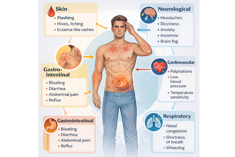
- Skin: flushing, hives, itching, eczema-like rashes.
- Gastrointestinal: bloating, diarrhoea, abdominal pain, and reflux.
- Neurological: headaches, dizziness, anxiety, insomnia, brain fog.
- Cardiovascular: palpitations, low blood pressure, temperature sensitivity.
- Respiratory: nasal congestion, shortness of breath, wheezing.
Because of this diversity, histamine-related conditions are often mistaken for food allergies, chronic fatigue, or anxiety disorders.
Histamine-Producing Gut Bacteria – and How to Rebalance Them
The gut microbiome contains both histamine-producing and histamine-degrading species. An imbalance in favour of histamine producers can worsen intolerance symptoms, even when diet is well-managed. Understanding which microbes contribute to excess histamine helps guide probiotic and nutritional choices.
Common Histamine-Producing Bacteria
- Lactobacillus reuteri – often found in probiotic supplements; can raise histamine levels in sensitive individuals.
- Lactobacillus casei – certain strains have been shown to stimulate histamine release in the gut under specific conditions.
- Lactobacillus bulgaricus – used in yoghurt production; may aggravate histamine symptoms.
- Enterococcus faecalis and Enterococcus faecium – natural intestinal residents that can overproduce histamine under stress or dysbiosis.
- Escherichia coli (certain strains) – capable of converting histidine into histamine during gut inflammation.
- Morganella morganii and Klebsiella pneumoniae – opportunistic bacteria linked with foodborne histamine toxicity (“scombroid” reactions).
Histamine-Degrading & Balancing Bacteria
- Bifidobacterium longum – supports gut barrier function and may reduce histamine-related inflammation.
- Bifidobacterium infantis – calms mast cell activation and promotes intestinal tolerance.
- Lactobacillus plantarum – shown to degrade histamine and strengthen mucosal integrity.
- Streptococcus thermophilus – neutral or mildly beneficial for histamine balance in many individuals.
Strategies to Rebalance the Microbiome
Eliminate or rotate probiotic strains that worsen symptoms, especially those containing L. reuteri or L. casei.
Introduce non-histamine-producing probiotics slowly to assess tolerance.
Support microbial diversity with fibre-rich foods (such as oats, flaxseed, and root vegetables).
Reduce gut inflammation with omega-3 fatty acids, quercetin, and polyphenol-rich foods like blueberries and green tea.
Address underlying dysbiosis through stool analysis or microbiome mapping if symptoms persist.
Tip: Changes in gut flora take time - consistent, gradual dietary and probiotic adjustments are more effective than rapid restriction or heavy supplementation.
References
– Maintz, L., & Novak, N. (2007). Histamine and histamine intolerance. American Journal of Clinical Nutrition, 85(5), 1185–1196.
– Comas-Basté, O. et al. (2020). Histamine intolerance: The current state of the art. Biomolecules, 10(8), 1181.
– Smolinska, S., Jutel, M., Crameri, R., & O’Mahony, L. (2014). Histamine and gut mucosal immune regulation. Allergy, 69(3), 273–281.
Gut Bacteria & Histamine Balance — Quick Reference
This table summarises common histamine producers, histamine degraders, and neutral/variable species. Individual responses vary: use as a guide alongside clinical judgement.
| Histamine Producers | Histamine Degraders / Balancers | Neutral / Variable (Context-Dependent) |
|---|---|---|
| Lactobacillus reuteri Lactobacillus casei Lactobacillus bulgaricus Enterococcus faecalis Enterococcus faecium Escherichia coli (some strains) Klebsiella pneumoniae Morganella morganii | Bifidobacterium longum Bifidobacterium infantis Lactobacillus plantarum Lactobacillus rhamnosus GG Akkermansia muciniphila (barrier support) Faecalibacterium prausnitzii (anti-inflammatory) | Streptococcus thermophilus Lactobacillus helveticus Lactococcus lactis Bacillus coagulans Saccharomyces boulardii (yeast; often supportive but individual variation) |
How to use: If symptoms worsen with a probiotic, check whether it includes strains in the “Producers” column. Trial alternatives from the “Degraders/Balancers” column and introduce gradually.
Notes: Effects are strain-specific and context-dependent (diet, inflammation, medications). Prioritise dietary fibre, polyphenols, sleep, and stress regulation to improve tolerance.
Dietary and Lifestyle Considerations
People with histamine intolerance often benefit from strategies that lower histamine load and support gut integrity:
- Reduce or avoid high-histamine foods such as aged cheeses, fermented products, smoked meats, alcohol, and leftovers.
- Eat freshly prepared foods and avoid long storage times, as histamine rises in ageing or spoilt food.
- Support DAO enzyme function with nutrients such as vitamin C, vitamin B6, copper, and magnesium.
- Consider probiotics that are non-histamine producing, such as Bifidobacterium infantis or Lactobacillus plantarum.
- Manage stress and ensure adequate sleep, as stress hormones can activate mast cells.
Low-Histamine Foods & Probiotic Support
Managing histamine load often starts with food choices and microbiome balance. A well-planned low-histamine approach can calm inflammation while supporting healthy enzyme function and gut ecology.
Foods Generally Low in Histamine
Freshly cooked meats and poultry (not aged, smoked, or processed)
Most fresh vegetables – especially courgette, carrots, pumpkin, and green beans
Gluten-free grains – rice, quinoa, oats, millet
Fresh fruits – apples, pears, blueberries, watermelon
Herbal teas – chamomile, rooibos, ginger
Healthy fats – olive oil, flaxseed oil, coconut oil
Tip: Prepare food fresh and avoid long storage times or leftovers, as histamine levels increase in aged or reheated foods.
Foods to Limit or Avoid
Aged cheeses, smoked meats, and tinned fish
Fermented foods (e.g., sauerkraut, kimchi, kombucha)
Alcohol, especially wine and beer
Tomatoes, spinach, aubergine, and avocado (histamine liberators)
Processed or pre-packaged meals
Microbiome & Probiotic Support
Choose probiotics that are non-histamine producing and support mucosal health:
Bifidobacterium infantis
Bifidobacterium longum
Lactobacillus plantarum
Lactobacillus rhamnosus GG (tolerated by many people)
Avoid probiotics that tend to raise histamine levels, such as Lactobacillus reuteri or Lactobacillus casei, unless guided by clinical advice.
Supportive Nutrients
Vitamin C – natural antihistamine and DAO co-factor
Vitamin B6 – supports DAO enzyme activity
Copper & Magnesium – involved in histamine metabolism
Omega-3 fatty acids – reduce inflammation and mast cell reactivity
Note: Always introduce dietary changes gradually and consult a qualified practitioner if you suspect MCAS or histamine intolerance. Over-restriction can reduce nutrient diversity and harm the microbiome in the long term.
Current Research Directions
Emerging studies explore how microbiome therapies might help regulate histamine activity:
- Targeted probiotics and postbiotics may stabilise mast cells and enhance gut barrier resilience.
- Low-histamine or anti-inflammatory diets show anecdotal success, but larger controlled trials are still limited.
- Future precision medicine approaches may use microbiome sequencing to identify bacterial strains associated with individual histamine reactivity.
When to Seek Medical Advice
Persistent or multisystem symptoms should be discussed with a clinician familiar with MCAS and histamine intolerance. Diagnosis is based on clinical history, response to low-histamine protocols, and sometimes laboratory markers such as DAO levels, plasma histamine, or tryptase.
Because symptoms overlap with many other conditions, professional evaluation is essential before starting restrictive diets or supplements. However, that said, many patients report disappointment with medical professionals' lack of knowledge and training in this area.
Systemic Mastocytosis
Systemic mastocytosis is a rare disorder characterised by an abnormal increase and accumulation of mast cells in multiple organs, most often the bone marrow, liver, spleen, skin, and gastrointestinal tract. Mast cells are immune cells that release histamine and other inflammatory mediators as part of the body’s defence system. When these cells proliferate excessively or become overactive, they can trigger widespread inflammatory and allergic-type reactions throughout the body.
The condition is classified as a form of mast cell disease, distinct from mast cell activation syndrome (MCAS). Whereas MCAS involves excessive activation of normal mast cells, systemic mastocytosis involves an actual overproduction of abnormal mast cells that often carry mutations in the KIT gene (most commonly KIT D816V). This mutation causes mast cells to grow and survive longer than usual.
Common symptoms arise from both the excess of mast cells and the substances they release. These can include flushing, itching, abdominal pain, diarrhoea, nausea, bone pain, fatigue, dizziness, low blood pressure, and sometimes anaphylaxis-like episodes. Because mast cells affect multiple systems, symptoms can mimic allergies, irritable bowel syndrome, or autoimmune conditions.
It should be noted that, due to the wide range of symptom variance and a lack of knowledge among most medical professionals, obtaining an accurate diagnosis can be a very frustrating experience that can take many years.
Diagnosis typically involves blood and urine tests for mast cell mediators such as tryptase, histamine, or prostaglandin D₂, along with a bone marrow biopsy to identify abnormal mast cells and confirm KIT mutations. The World Health Organisation (WHO) defines several subtypes, ranging from indolent forms (slow-growing and manageable) to aggressive systemic mastocytosis and mast cell leukaemia.
Treatment focuses on stabilising mast cells, reducing mediator release, and managing specific symptoms. Antihistamines (H₁ and H₂ blockers), mast cell stabilisers (such as cromolyn sodium or ketotifen), leukotriene inhibitors, and corticosteroids may be prescribed. For advanced or aggressive forms, targeted therapies such as tyrosine kinase inhibitors (e.g., midostaurin or avapritinib) are used to inhibit KIT-driven mast cell proliferation.
Systemic mastocytosis is a chronic condition requiring ongoing medical monitoring, usually under the care of a haematologist or immunologist. Lifestyle adjustments (such as avoiding known triggers (temperature extremes, alcohol, insect stings, stress, and certain medications)) can significantly reduce the frequency and intensity of reactions.
Histamine intolerance and mast cell activation reflect a wider immune–microbiome imbalance. A healthy gut ecosystem helps regulate inflammatory mediators, enzyme function, and immune tolerance. Through balanced nutrition, stress management, and careful microbiome support, many individuals find significant symptom improvement without reliance on long-term medication.
In restoring microbial balance, the aim is not to suppress the immune system but to help it respond appropriately - with precision rather than overreaction.
Peer-support community (non-medical)
Mast Cell Disease Group on Reddit
Histamine Intolerance, MCAS, Systemic Mastocytosis and Leukaemia
These four conditions exist along a spectrum of mast-cell-related and histamine-mediated disorders. Although their symptoms may overlap, they differ significantly in mechanism, pathology and clinical severity.
| Condition | Primary Mechanism | Key Features | Relationship to Others |
|---|---|---|---|
| Histamine Intolerance (HIT) | Functional overload of histamine relative to breakdown capacity (low DAO or HNMT activity). | Flushing, headaches, nasal congestion, hives, digestive upset, anxiety, and insomnia. Symptoms vary with diet and stress. | Not a mast-cell disorder. Often secondary to gut inflammation, dysbiosis, or nutrient deficiency. May mimic MCAS symptoms. |
| Mast Cell Activation Syndrome (MCAS) | Inappropriate release of histamine and other mediators from normal-number mast cells. | Episodic symptoms across multiple systems (skin, gut, cardiovascular, neurological). Triggered by stress, infection, or certain foods. | Mast cells are hyper-reactive but not proliferative. DAO deficiency may coexist, worsening histamine load. |
| Systemic Mastocytosis (SM) | Clonal proliferation of mast cells (often KIT D816V mutation) infiltrating bone marrow and other organs. | Mast cells are hyper-reactive but not proliferative. DAO deficiency may coexist, worsening the histamine load. | A rare myeloproliferative neoplasm. May present with MCAS-like symptoms but with abnormal mast-cell accumulation and mutation. |
| Leukaemia (esp. mast-cell or myeloid types) | Malignant transformation of bone marrow stem cells causing uncontrolled proliferation of immature white cells. | High serum tryptase, anaphylaxis, abdominal pain, bone lesions, and organ infiltration. | Very rare progression from systemic mastocytosis to mast-cell leukaemia. Most leukaemias do not involve mast cells. |
Summary Hierarchy:
Histamine Intolerance → Functional metabolic overload
MCAS → Hyperactive mast cells (normal number)
Systemic Mastocytosis → Neoplastic mast-cell proliferation
Leukaemia → Malignant marrow proliferation (may include mast-cell lineage)
Key Distinctions and Integrative Context
- Shared mediators: All can involve histamine release, but only mast-cell disorders (MCAS, SM, leukaemia) arise from mast-cell dysfunction or proliferation.
- DAO and gut health: Histamine intolerance is metabolic rather than neoplastic, often tied to intestinal inflammation or dysbiosis.
- Emotional and neurological effects: Mast-cell mediators affect the vagus nerve and central histamine receptors, influencing anxiety, fatigue, and sensory hypersensitivity—relevant in IEMT’s exploration of chronicity patterns.
- Diagnostic markers: HIT → low DAO, normal tryptase. MCAS → episodic tryptase rise. SM → persistently high tryptase and KIT mutation. Leukaemia → abnormal blast cells in marrow.
Clinical significance: Histamine intolerance and mild mast-cell activation are increasingly recognised in chronic fatigue, IBS, and anxiety-spectrum presentations. However, systemic mastocytosis and leukaemia are rare, serious haematological conditions managed by specialist haematology teams.
For IEMT practitioners: Understanding these distinctions helps contextualise clients’ reports of “histamine issues” and physiological hypersensitivity. It also emphasises the importance of differentiating functional metabolic sensitivities from true neoplastic disease when interpreting chronic symptom patterns.