







Melzack and Wall’s Gate-Theory of Pain
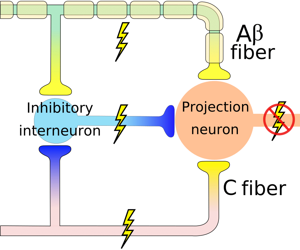
Melzack and Wall's "gate control theory of pain" is a pioneering model that explains how pain is perceived and modulated in the body. According to the theory, pain is not simply a direct result of tissue damage or injury, but rather is a complex process that involves multiple factors and mechanisms.
The theory was first proposed in 1965 by Ronald Melzack and Patrick Wall, who were researchers at McGill University in Canada. They proposed that pain perception is influenced by the activity of certain nerve fibres called "nociceptors," which are sensitive to tissue damage and transmit pain signals to the brain. However, they also recognized that other factors, such as the emotional and psychological state of the individual, can also influence pain perception.
According to the gate control theory, the perception of pain is mediated by a "gate" in the spinal cord, which can either amplify or inhibit the transmission of pain signals depending on the activity of other nerve fibres. For example, the activity of "large diameter" fibres, which transmit touch and pressure sensations, can inhibit the transmission of pain signals. This is why massaging a sore muscle or applying pressure to a wound can often provide relief from pain.
The gate control theory also proposes that the brain can exert a powerful influence on pain perception through various cognitive and emotional processes. For example, if an individual is distracted or engaged in a pleasurable activity, they may experience less pain than if they were focused on the sensation of pain.
In addition to explaining how pain is perceived and modulated, the gate control theory has also had significant practical implications for the treatment of pain. For example, it has helped to shed light on the effectiveness of certain pain management techniques, such as the use of non-pharmacological approaches like acupuncture and cognitive-behavioural therapy.
Overall, Melzack and Wall's gate control theory of pain is a highly influential model that has significantly impacted our understanding of pain and its perception. It has helped to shed light on the complex and multifaceted nature of pain and has had significant implications for the treatment of pain in clinical practice.

Nocioceptors
Pain receptors, also known as nociceptors, are specialized nerve endings that are responsible for detecting harmful stimuli, such as tissue damage or injury. These receptors are found throughout the body, including in the skin, muscles, joints, and internal organs.
When nociceptors are activated by harmful stimuli, they transmit a pain signal through sensory neurons to the spinal cord and brain. The pain signal is then processed and perceived by the brain, leading to the experience of pain.
There are several types of nociceptors that are activated by different types of stimuli. Some nociceptors are activated by mechanical stimuli, such as pressure or stretching, while others are activated by thermal stimuli, such as heat or cold. Still others are activated by chemical stimuli, such as irritants or inflammatory substances.
The activation of nociceptors is not always a reliable indicator of tissue damage or injury. In some cases, nociceptors may be activated by non-harmful stimuli, such as a light touch, due to abnormal activity or sensitivity. This can lead to the experience of chronic pain, which is pain that persists long after the original injury or damage has healed.
Pain Pathways
There are two main pathways for transmitting pain signals in the body: the spinal cord pathway and the brain pathway.
The spinal cord pathway involves the transmission of pain signals from nociceptors (pain receptors) in the body to the spinal cord, where they are processed and then transmitted to the brain. This pathway is responsible for providing a rapid response to pain, allowing us to quickly withdraw from harmful stimuli.
The brain pathway involves the transmission of pain signals from the spinal cord to the brain, where they are further processed and perceived as pain. This pathway is slower than the spinal cord pathway, but it allows for more complex processing of pain signals and can influence the emotional and cognitive responses to pain.
In addition to these two pathways, there are also several subcategories of nerve fibres that transmit pain signals. These include:
- A-delta fibres: These are medium-diameter fibres that transmit sharp, fast-acting pain signals.
- C fibres: These are small-diameter fibres that transmit slower, more diffuse pain signals.
- Adenosine triphosphate (ATP) fibres: These are fibres that transmit pain signals in response to chemical stimuli, such as inflammatory substances.
- Endogenous opioid fibres: These are fibres that release endogenous opioids, such as endorphins, in response to pain. Endogenous opioids can act as natural painkillers and help to modulate pain perception.
The transmission of pain signals in the body involves complex and multifaceted pathways that involve various types of nerve fibers and brain regions. Understanding these pathways can help to improve the diagnosis and treatment of pain conditions.
A-Delta Fibres
A-delta fibres, also known as Aδ fibres, are a type of sensory nerve fibre that transmits pain signals from the body to the central nervous system (CNS). These fibres are classified as "small diameter" fibres and are intermediate in size between large diameter fibres, such as myelinated A-beta fibres, and small diameter fibres, such as unmyelinated C fibres.
A-delta fibres are responsible for transmitting sharp, fast-acting pain signals, such as those that occur with a cut or a burn. They are activated by mechanical or thermal stimuli and are most sensitive to stimuli that are high in intensity or duration.
A-delta fibres are distinguished from other types of fibres by the presence of myelin, which is a fatty substance that surrounds the axon and helps to insulate it. Myelination allows A-delta fibres to transmit signals faster than C fibres, which are unmyelinated.
A-delta fibres are found throughout the body and are especially prevalent in the skin, where they are responsible for transmitting fast-acting pain signals that allow us to quickly withdraw from harmful stimuli. They are also found in other organs, such as the bladder and the intestine, where they help to detect pain and discomfort.
Overall, A-delta fibres play a crucial role in detecting and transmitting fast-acting pain signals in the body, helping to protect us from harm and alert us to potential injuries. Understanding the function and properties of A-delta fibres can help to improve the diagnosis and treatment of pain conditions.
C-Fibres
C-fibres, are a type of sensory nerve fibre that transmits pain signals from the body to the central nervous system (CNS). These fibres are classified as "small diameter" fibres and are distinguished from other types of fibres by their lack of myelin, which is a fatty substance that surrounds the axon and helps to insulate it.
C fibres are responsible for transmitting slow, diffuse pain signals, such as those that occur with chronic pain or inflammation. They are activated by mechanical, thermal, or chemical stimuli and are most sensitive to stimuli that are low in intensity or duration.
C fibres are found throughout the body and are especially prevalent in the skin, where they are responsible for transmitting slow, diffuse pain signals that allow us to detect and respond to more subtle or chronic pain. They are also found in other organs, such as the bladder and the intestine, where they help to detect pain and discomfort.
In addition to transmitting pain signals, C fibres also play a role in the immune response, releasing substances that can contribute to inflammation and swelling.
C fibres play a crucial role in detecting and transmitting slow, diffuse pain signals in the body, helping to alert us to potential injuries and chronic pain conditions. Understanding the function and properties of C fibres can help to improve the diagnosis and treatment of pain conditions.
Adenosine triphosphate (ATP) fibres
Adenosine triphosphate (ATP) fibres are a type of sensory nerve fibre that transmit pain signals in response to chemical stimuli, such as inflammatory substances. These fibres are activated by the release of ATP, which is a molecule that plays a central role in energy metabolism in cells.
ATP fibres are found throughout the body and are especially prevalent in the skin, where they help to detect and transmit pain signals in response to chemical stimuli. They are also found in other organs, such as the bladder and the intestine, where they help to detect pain and discomfort.
The activation of ATP fibres can contribute to the experience of pain, especially in the presence of inflammation or tissue damage. In addition to transmitting pain signals, ATP fibres can also release substances that can contribute to inflammation and swelling.
ATP fibres play a crucial role in detecting and transmitting pain signals in response to chemical stimuli in the body. Understanding the function and properties of ATP fibres can help to improve the diagnosis and treatment of pain conditions.
Endogenous opioid fibres
Endogenous opioid fibres are a type of nerve fibre that release endogenous opioids, such as endorphins, in response to pain. Endogenous opioids are natural painkillers that are produced by the body and act on opioid receptors to reduce the perception of pain.
Endogenous opioid fibres are found throughout the body and are especially prevalent in the brain and spinal cord, where they are involved in the modulation of pain perception. When activated, these fibres release endogenous opioids, which bind to opioid receptors and reduce the transmission of pain signals.
The activation of endogenous opioid fibres can provide natural pain relief and help to modulate pain perception. This is why activities such as exercise or laughter can sometimes provide relief from pain.
Endogenous opioids can also be used clinically to treat pain. Opioid medications, such as morphine and oxycodone, act on the same opioid receptors as endogenous opioids and can provide relief from severe or chronic pain.
Overall, endogenous opioid fibres play a crucial role in modulating pain perception through the release of endogenous opioids. Understanding the function and properties of these fibres can help to improve the diagnosis and treatment of pain conditions.