







Vagal Tone and Measurement
Vagal Tone and Measurement
Vagal tone refers to the activity of the vagus nerve: the primary parasympathetic pathway connecting the brain to the heart, lungs, and digestive system. It reflects the nervous system’s capacity to regulate physiological states flexibly and adaptively. High vagal tone is associated with calm alertness, emotional stability, and resilience under stress, whereas low vagal tone correlates with anxiety, inflammation, fatigue, and difficulties in emotional regulation.
In the context of Polyvagal Theory and therapeutic practice, understanding vagal tone provides insight into how clients’ autonomic systems respond to internal and external stimuli, offering a physiological dimension to emotional and behavioural assessment.
What Is “Vagal Tone”?
Vagal tone represents the level of parasympathetic influence exerted by the vagus nerve on the heart and other organs. It reflects how effectively the nervous system can transition between activation (sympathetic arousal) and relaxation (parasympathetic recovery). In simple terms, it measures the body’s ability to return to calm after stress. Vagal tone develops across early life through attuned caregiving, safety, and repeated regulation experiences and is commonly disrupted following chronic stress, developmental adversity, or trauma.
- High vagal tone: Indicates efficient heart–brain communication, flexible emotional responses, and greater physiological stability. Individuals typically recover from stress quickly and maintain social engagement.
- Low vagal tone: Suggests diminished parasympathetic regulation, often linked to chronic stress, trauma, inflammation, and mood disorders.
Because the vagus nerve modulates both heart rate and inflammation, vagal tone serves as a biomarker for integrated physical and psychological health. Approximately 80–90% of vagal fibres are afferent, carrying information from the body to the brain, which explains why the tone reflects bodily state awareness more than motor output.
Heart Rate Variability (HRV) as an Indirect Marker
Heart Rate Variability (HRV) (the variation in time between successive heartbeats) is widely used as a non-invasive measure of vagal tone. Rather than measuring heart rate itself, HRV examines how heart rhythm fluctuates in response to breathing, stress, and relaxation.
- High HRV: Reflects strong vagal modulation and adaptability. Seen in individuals with good emotional regulation, cardiovascular health, and resilience.
- Low HRV: Indicates reduced vagal control and rigidity in autonomic responses. Low HRV is often linked to conditions such as anxiety, depression, chronic pain, and inflammatory disorders.
HRV can be measured through wearable sensors or biofeedback devices using electrocardiography (ECG) or photoplethysmography (PPG). Frequency-domain analyses identify components such as the high-frequency (HF) band, which primarily reflects parasympathetic respiratory-linked vagal activity while remaining influenced by breathing pattern, posture, and overall autonomic balance.
However, HRV is an indirect measure - it reflects the balance of sympathetic and parasympathetic inputs rather than vagal activity alone. Interpretation should therefore be cautious, especially in clinical or therapeutic contexts.
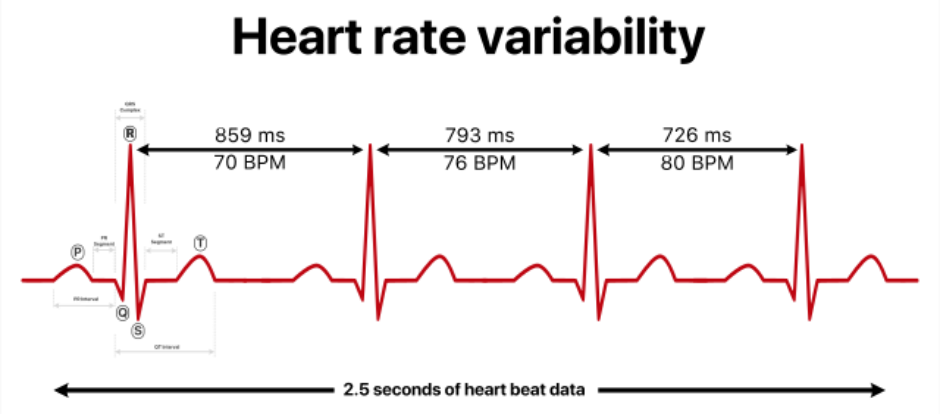
Adapted from: YitzhakNat. (n.d.). Heart rate variability (HRV) [SVG image]. Wikimedia Commons.
.svg)
License: CC BY-SA 4.0
Photoplethysmography (PPG)
Photoplethysmography (PPG) is a simple, non-invasive optical technique used to measure changes in blood volume within the microvascular tissue. It works by shining a small light - usually infrared or red - through the skin (commonly at the fingertip, earlobe, or wrist) and detecting how much light is absorbed or reflected by the blood as it pulses through the vessels.
Each heartbeat causes a slight change in blood volume, which alters light absorption. These fluctuations create a waveform known as a plethysmogram. By analysing this signal, PPG devices can estimate heart rate, respiration rate, and even heart rate variability (HRV), a key marker of vagal tone and autonomic balance.
Common Uses:
- Monitoring heart rate and pulse rhythm in wearable devices (e.g., smartwatches, fitness trackers)
- Estimating HRV as an indirect indicator of vagal activity and emotional regulation
Assessing blood oxygen saturation (SpO₂) in clinical and wellness settings
PPG is less precise than electrocardiography (ECG) but offers a convenient, accessible method for tracking autonomic patterns in daily life. Movement, temperature, and poor sensor contact can affect accuracy, so readings are best interpreted as trend indicators rather than diagnostic measurements.
For therapists and researchers interested in vagal tone, PPG provides a practical tool for observing real-time physiological changes during breathwork, relaxation, or emotional regulation exercises.
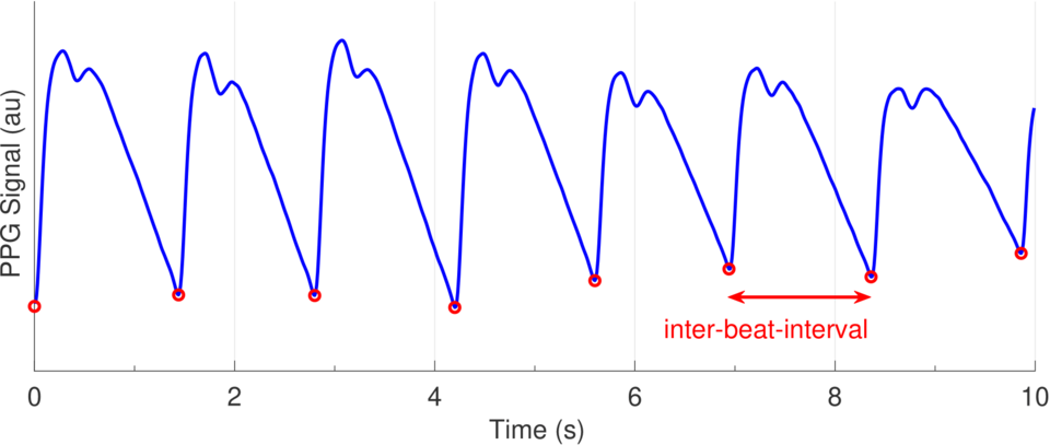
Adapted from: Charlton, P. H., Celka, P., Farukh, B., Chowdhury, M. F., & Hu, X. (2022). Acquiring wearable photoplethysmography data in daily life: The PPG Diary Pilot Study. Proceedings of the 7th International Electronic Conference on Sensors and Applications.
License: CC BY 4.0.
Breathwork, Posture, and Body Awareness in Assessing Vagal Responses
Vagal tone is not static; it can shift moment to moment depending on posture, breathing, and internal state. Therapists and practitioners can observe and influence vagal responses through embodied awareness techniques.
- Breathwork: Slow, rhythmic breathing (around 5–7 breaths per minute) enhances vagal afferent signalling via the nucleus tractus solitarius, increasing HRV and promoting calm.
- Posture: Upright, open body posture is commonly associated with more efficient parasympathetic engagement, while collapsed or constricted postures often accompany dorsal vagal shutdown and reduced autonomic flexibility.
- Facial and vocal tone: Soft prosody, gentle eye contact, and a relaxed facial expression reflect ventral vagal activation and can serve as real-time indicators of safety or threat responses.
- Interoceptive awareness: Mindful attention to heartbeat, breath, and internal sensations improves self-regulation and helps clients recognise when their nervous system is shifting state.
These physiological markers complement therapeutic observation, offering valuable insights into how emotional states manifest in the body.
Breathing Techniques to Improve Vagal Tone
Controlled breathing is one of the most effective and accessible ways to stimulate the vagus nerve and enhance parasympathetic regulation. Slow, rhythmic breathing strengthens the connection between the heart and the brain through vagal afferents, increasing heart rate variability (HRV) and promoting emotional calm. Below are three evidence-based methods that support vagal activation and autonomic balance.
1. Resonant (Coherent) Breathing
Method: Breathe at a steady pace of approximately 5–6 breaths per minute (about 5 seconds inhalation, 5 seconds exhalation). Continue for 5–10 minutes.
Effects: This breathing rhythm synchronises heart rate and respiration, maximising HRV and vagal engagement. It is used in biofeedback training and clinical stress reduction programmes.
2. Extended Exhalation Breathing
Method: Inhale gently through the nose for 4 seconds, then exhale slowly and completely through the mouth or nose for 6–8 seconds. Pause briefly before the next inhale.
Effects: Lengthening the exhalation stimulates efferent vagal fibres, activating the parasympathetic system. This technique is particularly advantageous for reducing anxiety, lowering heart rate, and calming the body before sleep or therapy sessions.
3. Box Breathing (Square Breathing)
Method: Inhale for 4 seconds, hold the breath for 4 seconds, exhale for 4 seconds, and hold again for 4 seconds. Repeat for several minutes.
Effects: Used by athletes, musicians, and clinicians, this technique enhances focus, stabilises the nervous system, and promotes mindful breathing control. The brief holds help integrate sympathetic and parasympathetic rhythms, improving overall vagal tone.
Additional Tips:
- Use nasal breathing where possible; it naturally slows respiration and supports nitric oxide production.
- Combine breathing with gentle movement or posture awareness to reinforce embodied calm.
- Practice in a quiet environment with relaxed shoulders and an open posture to facilitate ventral vagal activation.
- Regular practice of slow, deliberate breathing not only improves physiological regulation but also strengthens emotional resilience, making it a cornerstone of vagal toning and self-regulation strategies.
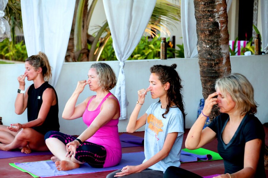
Each breath gently lowers arousal, signalling safety and restoring physiological balance.
Adapted from: Pranayama in Goa [Photograph] by renatoyoga. Wikimedia Commons. CC BY-SA 4.0

Ethical Considerations in Physiological Measurement for Therapists
Although HRV and other physiological tools can enrich therapeutic understanding, their use requires clear ethical boundaries. In some modalities (i.e., "Thought Field Therapy (TFT)"), it is not uncommon for practitioners to make claims about improving HRV and claim to measure this for their clients. It has been observed that some practitioners do not actually understand what HRV is and measure it using substandard tools.
All therapists must respect client autonomy, privacy, and data security and ensure that any such physiological assessments are appropriate to their scope of practice.
- Informed consent: Clients should fully understand what is being measured, why, and how their data will be used or stored.
- Non-diagnostic use: HRV and biofeedback readings should be used to support self-awareness, not for medical diagnosis or to predict pathology.
- Confidentiality: Physiological data are personal health information and should be protected under GDPR and data protection laws.
- Competence: Practitioners must have appropriate training to interpret physiological metrics responsibly and avoid overgeneralisation.
Used ethically, physiological awareness and vagal monitoring can deepen therapeutic insight into clients’ regulation capacities, enhance safety in trauma work, and foster a more embodied understanding of psychological healing.