






The Vagal Branches: Cardiac, Pulmonary, Gastrointestinal, Auricular
Overview
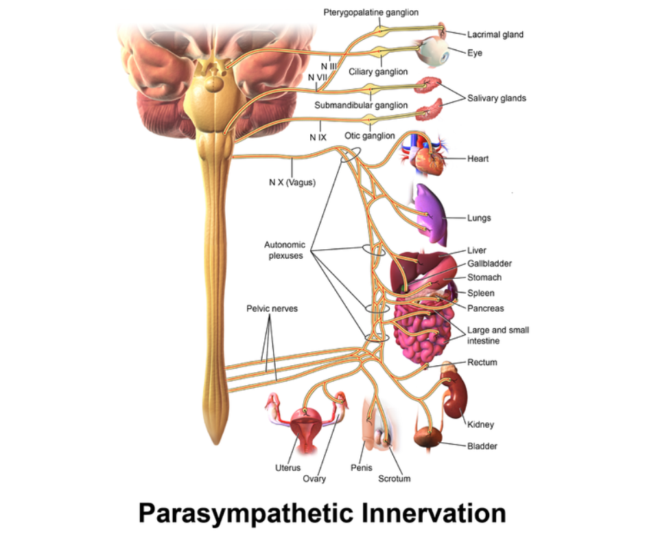
The vagus nerve branches extensively as it descends from the brainstem, forming a network that supplies the heart, lungs, digestive tract, and parts of the ear. Each branch serves distinct yet interconnected physiological functions that collectively maintain homeostasis and link internal regulation to emotional experience. Through these pathways, the vagus nerve modulates vital processes such as heart rhythm, respiration, digestion, and even aspects of communication and social engagement.
The complexity of these branches reflects the vagus nerve’s integrative role across multiple organ systems. While each division performs specialised tasks, they operate together within the broader autonomic network, responding dynamically to cues of safety, stress, and internal need.
Homeostasis
'Homeostasis' refers to the body’s ability to maintain stable internal conditions despite external changes. It involves continuous regulation of variables such as temperature, pH, blood pressure, and glucose levels to keep them within a narrow physiological range. This stability is achieved through feedback mechanisms coordinated by the nervous and endocrine systems, particularly the hypothalamus and autonomic pathways.
The vagus nerve plays a central role in homeostatic regulation by modulating heart rate, digestion, respiration, and immune activity. Through constant afferent and efferent signalling, it helps balance energy use, recovery, and adaptation, ensuring that internal processes remain aligned with the body’s environmental and emotional demands.
Cardiac Branches
The cardiac branches of the vagus nerve originate primarily from the nucleus ambiguus in the medulla oblongata and project to the heart via the cardiac plexus. These fibres provide parasympathetic input to the sinoatrial (SA) and atrioventricular (AV) nodes, which regulate the heartbeat. Activation of these fibres slows the heart rate, reduces cardiac output, and promotes energy conservation. Cardiac vagal afferents also transmit baroreceptor feedback related to blood pressure, allowing rapid moment-to-moment regulation of cardiovascular stability in response to posture, effort, and emotional state.
This vagal control of the heart forms the basis of respiratory sinus arrhythmia, the natural fluctuation in heart rate that occurs during breathing. The phenomenon reflects healthy vagal tone and flexibility in the autonomic nervous system. Reduced vagal activity, by contrast, is associated with emotional dysregulation, chronic stress, and cardiovascular strain.
Sinoatrial (SA) and Atrioventricular (AV) Nodes
The sinoatrial (SA) and atrioventricular (AV) nodes are specialised clusters of cardiac muscle cells that coordinate the heart’s electrical activity. Together, they ensure the heart contracts rhythmically and efficiently, maintaining consistent blood flow throughout the body.
The SA node, located in the right atrium, acts as the heart’s natural pacemaker. It generates rhythmic electrical impulses that initiate each heartbeat and set the overall pace of cardiac activity. These impulses spread across the atria, causing them to contract and push blood into the ventricles.
The AV node, situated between the atria and ventricles, receives and briefly delays the electrical signal from the SA node before transmitting it to the ventricular conduction system. This delay allows the ventricles to fill before contraction. Parasympathetic input from the vagus nerve modulates both nodes, slowing the heart rate during rest and enhancing energy efficiency.
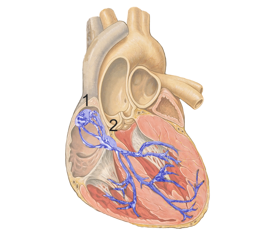
Source: Adapted from Heart (anterior view, coronal section), illustrated by Patrick J. Lynch, MD, with C. Carl Jaffe, MD, Yale University Center for Advanced Instructional Media.

Pulmonary Branches
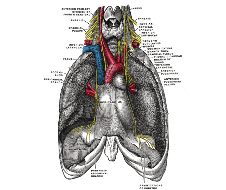
The pulmonary branches arise from both the right and left vagus nerves and enter the lungs via the pulmonary plexuses. These branches innervate the bronchial tree and visceral pleura, regulating the constriction and dilation of airways and modulating the rhythm of respiration. They also participate in protective reflexes such as coughing and bronchoconstriction in response to irritants. Pulmonary vagal afferents further relay chemoreceptor information related to oxygen and carbon dioxide levels, directly linking respiratory chemistry to autonomic and emotional regulation.
Through these pathways, the vagus nerve contributes to the coupling between breathing and emotional state. Slow, controlled breathing enhances vagal activity, promoting relaxation and parasympathetic dominance. This physiological link underlies many calming and grounding practices, in which breathing influences the body’s internal state.
The Visceral Pleura
The visceral pleura is the delicate membrane that directly covers the surface of the lungs. It forms a continuous layer that folds back at the lung roots to become the parietal pleura, which lines the inner chest wall. Together, these two layers create the pleural cavity, a thin fluid-filled space that allows the lungs to move smoothly during breathing.
The visceral pleura contains sensory and autonomic nerve fibres, blood vessels, and lymphatics that help maintain pulmonary function and responsiveness. Through its connections with the vagus nerve and pulmonary branches, it contributes to the reflex regulation of respiration and the detection of mechanical changes in the lungs.
Gastrointestinal Branches
The gastrointestinal branches form one of the most extensive divisions of the vagus nerve, supplying the oesophagus, stomach, pancreas, and intestines. These fibres coordinate digestive processes, including peristalsis, secretion of digestive enzymes, and regulation of gastric acid production. They also carry afferent sensory information from the gut to the brain about nutrient content, satiety, and mechanical stretch.
Through this continual communication, the gastrointestinal branches play a key role in the gut–brain axis. They mediate the gut microbiota's influence on brain function and emotional regulation. Vagal afferents detect inflammatory mediators, cytokines, and metabolic by-products produced by gut bacteria, relaying this information to the brainstem and limbic regions to shape immune responses, mood, and stress reactivity. This feedback system ensures that digestion, immunity, and mood remain tightly coordinated.
Auricular Branch (Arnold’s Nerve)
The auricular branch, or Arnold’s nerve, is the only cutaneous branch of the vagus nerve. It provides sensory innervation to parts of the external ear and ear canal. Though small, it demonstrates the vagus nerve’s wide distribution and its integration of external sensory input with internal regulation.
Stimulation of this branch (such as through gentle touch, sound vibration, or non-invasive transcutaneous vagal nerve stimulation (tVNS)) has been shown in research to activate central vagal pathways and influence parasympathetic tone. This connection between tactile sensation and autonomic response reflects how even subtle sensory cues can engage the body’s regulatory systems, linking physical contact, sound, and emotional calm through shared neural pathways.
A Curious Anecdote
The auricular branch of the vagus nerve has occasionally been nicknamed the Alderman’s nerve, a title linked to a curious old social custom. During formal banquets, it was noted that some guests (particularly elderly city aldermen) would cough, gag, or even faint when their ears were tickled or cleaned.
A popular anecdote, repeated in several anatomical sources, suggests that the name arose from a ceremonial practice among London aldermen, who placed rosewater-soaked napkins behind their ears during feasts, believing this aided digestion.
The underlying mechanism is physiological rather than mystical: this branch of the vagus nerve provides sensory innervation to part of the external ear canal. When stimulated, it can evoke the ear-cough reflex (also called Arnold’s reflex), activating the vagus nerve and producing coughing or other parasympathetic effects, hence the enduring moniker, “Alderman’s nerve”.
Functional Integration
The cardiac, pulmonary, gastrointestinal, and auricular branches form a unified system that maintains vital bodily rhythms and underpins many aspects of emotional life. By adjusting heart rate, respiration, and digestion, the vagus nerve ensures that the body remains responsive yet stable across changing circumstances. These mechanisms operate automatically but are profoundly influenced by emotional states, perceptions of safety, and interpersonal context, the very domains in which IEMT practitioners observe shifts during therapeutic work as nervous system regulation reorganises.